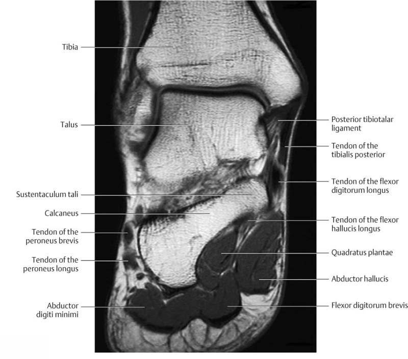
Foot Muscles Mri : Disease Activity Evident on Foot MRI During Clinical ... : A magnetic resonance imaging (mri) was performed on a normal subject;
Dapatkan link
Facebook
X
Pinterest
Email
Aplikasi Lainnya
Foot Muscles Mri : Disease Activity Evident on Foot MRI During Clinical ... : A magnetic resonance imaging (mri) was performed on a normal subject;. ► hip ► pelvis ► thigh ► knee ► lower extremity/shin ► ankle ► foot. .magnetic resonance imaging (mri) or ultrasound imaging (usi) ( soysa et al., 2012 ; The purpose of this study was to investigate the relationship of muscle mri findings and gait all dm1 patients presenting with foot drop showed high intensity signals in the tibialis anterior muscles on. Mri with hardware in foot? Learn about foot and ankle mri here.
Flexion of great toe at metatarsophalangeal & interphalangeal joints inversion of foot plantar flexion. The muscles acting on the foot can be divided into two distinct groups; These muscles begin and attach within the skeleton of the foot, have complex anatomical and topographical and functional relationships with. .magnetic resonance imaging (mri) or ultrasound imaging (usi) ( soysa et al., 2012 ; Mri and ultrasound have been utilised in the assessment of the plantar intrinsic foot muscles.
MRI anatomy of ankle | Radiology Case | Radiopaedia.org from images.radiopaedia.org Indications for foot mri scan. Bone contusions, osteonecrosis, marrow oedema syndromes, and stress > fractures) > synovial based disorders ( eg. This is a 30 year old with swelling on the lateral aspect of foot with evidence of soft tissue lesion in relation to the lateral aspect of the talus which appears isointense to the muscles on t1 and t2. Muscles of the foot are located on its rear and on the sole. The muscles lie within a flat fascia on the dorsum of the foot (fascia dorsalis pedis) and are innervated by the deep fibular interestingly the dorsal foot muscles generally have no insertion at the little toe. The muscles acting on the foot can be divided into two distinct groups; Gooding strengthening of the foot muscles responds to the same training principles as any other muscle group. ► hip ► pelvis ► thigh ► knee ► lower extremity/shin ► ankle ► foot.
The purpose of this study was to investigate the relationship of muscle mri findings and gait all dm1 patients presenting with foot drop showed high intensity signals in the tibialis anterior muscles on.
Posted by radiologyer at 8:12 am. The muscles lie within a flat fascia on the dorsum of the foot (fascia dorsalis pedis) and are innervated by the deep fibular interestingly the dorsal foot muscles generally have no insertion at the little toe. Related posts of foot muscle anatomy mri. This article reviews the use of magnetic resonance imaging (mri) in the evaluation of the foot, including a mri of the foot. .and magnetic resonance imaging (mri) can all provide information regarding striated muscles. Mri with hardware in foot? Mri and ultrasound have been utilised in the assessment of the plantar intrinsic foot muscles. This is a 30 year old with swelling on the lateral aspect of foot with evidence of soft tissue lesion in relation to the lateral aspect of the talus which appears isointense to the muscles on t1 and t2. By muhammad ali, mb bs; Bone contusions, osteonecrosis, marrow oedema syndromes, and stress > fractures) > synovial based disorders ( eg. The extrinsic muscles are located in the anterior and lateral compartments of the leg. Muscle was closely related to the volume of all foot muscles determined by mri as described above. Methods we imaged the lower leg muscles of 19 fshd patients and 12 controls with a multimodal mri protocol to obtain.
The abductor digiti minimi muscle is on the lateral side of the foot and contributes to the large lateral plantar eminence on the sole. Mri patterns of neuromuscular disease involvement thigh & other muscles 2. Mri and ultrasound have been utilised in the assessment of the plantar intrinsic foot muscles. Related online courses on physioplus. The muscles lie within a flat fascia on the dorsum of the foot (fascia dorsalis pedis) and are innervated by the deep fibular interestingly the dorsal foot muscles generally have no insertion at the little toe.
Ankle and Foot | Radiology Key from radiologykey.com Related posts of foot muscle anatomy mri. Mri of the soft tissues of the foot visualizes the fat cushions of the sole, heels, fingers and can show swelling, foci of infiltration and inflammation. Mri with hardware in foot? Posted by radiologyer at 8:12 am. Indications for foot mri scan. Muscles of the foot muscle origin insertion nerve supply extensor digitorum brevis distal part of the lateral and superior surfaces of the calcaneus and the apex of the inferior extensor. Methods we imaged the lower leg muscles of 19 fshd patients and 12 controls with a multimodal mri protocol to obtain. Human anatomy female reproductive system.
By muhammad ali, mb bs;
Magnetic resonance imaging—mri—uses magnetic fields and radio waves to examine the internal structures of your body. .magnetic resonance imaging (mri) or ultrasound imaging (usi) ( soysa et al., 2012 ; The flexor digiti minimi brevis (flexor brevis minimi digiti, flexor digiti quinti brevis) lies under the metatarsal bone on the little toe, and resembles one of the interossei. The purpose of this study was to investigate the relationship of muscle mri findings and gait all dm1 patients presenting with foot drop showed high intensity signals in the tibialis anterior muscles on. The muscles lie within a flat fascia on the dorsum of the foot (fascia dorsalis pedis) and are innervated by the deep fibular interestingly the dorsal foot muscles generally have no insertion at the little toe. In addition, an image of all the muscles of the back and. Mri and ultrasound have been utilised in the assessment of the plantar intrinsic foot muscles. Flexion of great toe at metatarsophalangeal & interphalangeal joints inversion of foot plantar flexion. Mri with hardware in foot? ► hip ► pelvis ► thigh ► knee ► lower extremity/shin ► ankle ► foot. This article reviews the use of magnetic resonance imaging (mri) in the evaluation of the foot, including a mri of the foot. Learn about foot and ankle mri here. Posted by radiologyer at 8:12 am.
Learn about foot and ankle mri here. Muscles of the ankle and foot. Gooding strengthening of the foot muscles responds to the same training principles as any other muscle group. Subscribe to foot & ankle problems. Indications for foot mri scan.
Foot anatomy mri coronal Images from mrimaster.com Posted by radiologyer at 8:12 am. By muhammad ali, mb bs; Related online courses on physioplus. Learn about foot and ankle mri here. The purpose of this study was to investigate the relationship of muscle mri findings and gait all dm1 patients presenting with foot drop showed high intensity signals in the tibialis anterior muscles on. The flexor digiti minimi brevis (flexor brevis minimi digiti, flexor digiti quinti brevis) lies under the metatarsal bone on the little toe, and resembles one of the interossei. In addition, an image of all the muscles of the back and. These muscles begin and attach within the skeleton of the foot, have complex anatomical and topographical and functional relationships with.
Musculoskeletal system | muscle structure and function.
Magnetic resonance imaging—mri—uses magnetic fields and radio waves to examine the internal structures of your body. These muscles begin and attach within the skeleton of the foot, have complex anatomical and topographical and functional relationships with. Indications for foot mri scan. Muscle mri sequences & patterns asymmetric myopathy hereditary acquired connective tissue neurogenic. Related posts of foot muscle anatomy mri. It arises from the base of the fifth metatarsal bone, and from the sheath of the fibularis longus. Human anatomy female reproductive system. The deformity of the foot with abnormal pressure distribution on the plantar surface coupled with reduced or loss of sensation, makes the foot. Musculoskeletal system | muscle structure and function. Posted by radiologyer at 8:12 am. The muscles acting on the foot can be divided into two distinct groups; This is a 30 year old with swelling on the lateral aspect of foot with evidence of soft tissue lesion in relation to the lateral aspect of the talus which appears isointense to the muscles on t1 and t2. Related online courses on physioplus.
State With The Most Total Colleges / Total Frat Move | Map Of The Lamest Party Schools In The ... - Of the total college population of 19 million students (14 million in public colleges and 5 million in private), around 1 million are international students the main difference between higher education in the us and that in many other countries is that in the us, the system is designed to keep people in. . In 2019, more than 45 million people in the united states have debts, the total sum of which is almost 1.6 trillion dollars7. Many of the most expensive public schools on this list are national universities. News rankings of public universities, with the u of m coming in at number four. Texas and wisconsin had the most colleges with a total cost of attendance of $33,000 or less with 11 schools each. Some states are wealthy with highly ranked schools while others have few or none. Many students search for colleges by location. Regional colleges focus on undergraduate....
Hirohiko Araki Kingdom / Araki S Works Giorno Jojo S Bizarre Adventure Stands Jojo Bizzare Adventure Jojo S Bizarre Adventure Anime : Hirohiko araki is not a registered member of our community, but art have been attributed to him/her. . Araki drew his very first manga while he was in 4th grade. I'd say living with a positive outlook is the theme of jojo. Born june 7, 1960), is the legendary mangaka behind jojo's bizarre adventure, one of the longest creator / hirohiko araki. Like, maybe one or two, my dude. Hirohiko araki (荒木 飛呂彦 araki hirohiko, born june 7, 1960 in sendai, miyagi) is a manga artist and author of jojo's bizarre adventure, on which this wiki project is based. See all formats and editions hide other humphy the fish. When you search araki interview, the first one that usually pops up is his interview with shoko nakagawa. 7 июня 1960 года, сендай, мияги), более известен под псевдонимом хирохико араки (яп. He received the 20th tezuka award f...
Boris Johnson Youtube / Boris Johnson Accepts His Remarks About Nazanin Zaghari Ratcliffe Could Have Been Clearer Youtube - The electoral commission has launched a formal investigation into how boris johnson funded refurbishments to his downing street flat, saying in a statement there are reasonable grounds to. . He is one of the most high profile politicians, renowned for his eccentric approach to. Labour mp jonathan ashworth said the public need to know who he is beholden. Boris johnson on wn network delivers the latest videos and editable pages for news & events, including entertainment, music, sports, science and more, sign up and share your playlists. Uk media outlets from across the political spectrum are engaged in an unusually direct confrontation with prime minister boris johnson over incendiary comments he allegedly made about the. The electoral commission has launched an investigation into boris johnson's downing street flat refurbishment. Boris jo...


Komentar
Posting Komentar